Understanding the Complexity of the Human Heart with 3D Bioprinting
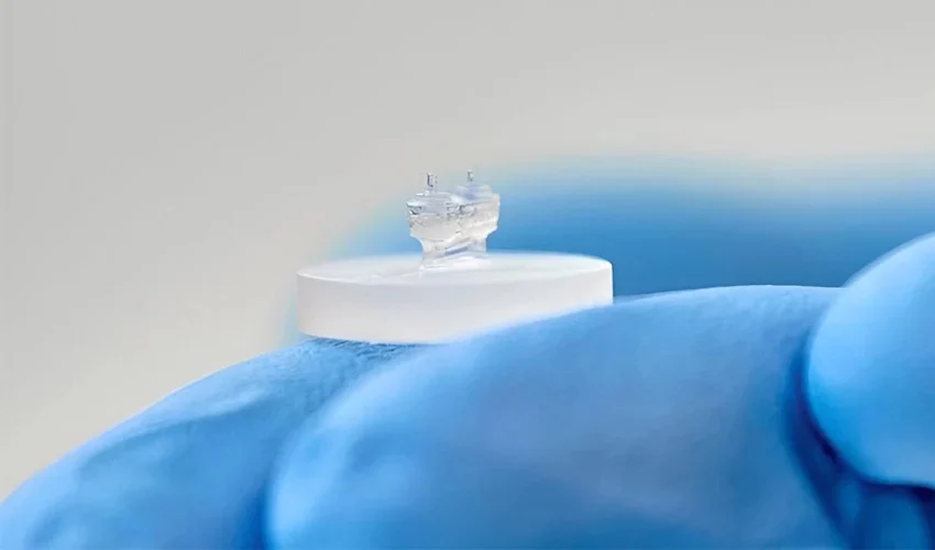
The heart is a complex organ, with mechanics and physiology that are difficult to study and understand. However, 3D printing could help us to better understand this vital part of our body. In any case, this is the aim of a team of researchers from the Azrieli Center at CHU Sainte-Justine in Canada. They have developed a “heart-on-a-chip”: a system made using 3D bioprinting that simulates the mechanical and electrical activity of the human heart, thus mimicking its behavior. The device could make it easier to study individual heart diseases and develop specific treatments more suited to each patient.
For some years now, 3D bioprinting, which involves printing with living cells, has been evolving rapidly, and has been the driving force behind a number of promising and encouraging medical projects. Ultimately, one of the goals of those using this technology would be to develop viable organs, thus considerably reducing the number of people waiting for a kidney, liver or heart. Transplants could be faster and potentially less risky. However, there is still a long way to go before we get there, still bioprinting promises great medical advances. For the time being, it is mainly used in research phases, offering alternative solutions to better understand a given disease.
The process developed by the Canadian researchers
Using 3D Bioprinting to Study the Human Heart
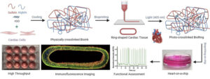
Notably, diseases affecting the heart are particularly complex. Because the heart is such a vital organ, it is currently very difficult for medical professionals to study its cellular activity live and draw conclusive conclusions. That’s why researchers at CHU Sainte-Justine have set out to create a bio-printed ring that reproduces the physiology and mechanics of the heart. They developed a specific bio-ink based on photocrosslinkable natural polymers, methacryloyl gelatin (GelMA), methacrylate alginate (AlgMA), and electroconductive nanomaterials of reduced graphene oxide (rGO). This combination resulted in a material that was not only easier to print, but also incorporated improved tensile and compressive mechanical properties.
Researchers have therefore 3D printed heart tissue in the shape of a ring, making it easier to study the organ’s behavior. It’s a kind of chip that can be more easily produced and analyzed. Houman Sovaji, Professor of Pharmacology and leader of the study, explains: “Our research has made it possible to combine 3D bioprinting technology to produce standard hearts-on-a-chip much more quickly and precisely. What’s more, our results show that printed devices perform better than those produced manually.”
Ultimately, the researchers hope to be able to use the patient’s stem cells to develop models of heart disease and test treatments directly on the chip and therefore on the cells. This opens up new prospects for the creation of more effective, personalized drugs.
Ali Mousavi, another author of the study, concludes, “The next step will be to compare healthy and diseased heart cells to develop solid cardiac pathology models. That will also let us safely and accurately test the effect of new therapeutic molecules on cells.” In the meantime, you can access the full study HERE.
*All Photo Credits: Centre Azrieli/CHU Sainte-Justine